====
Good Hope Hospital Eye Clinic

Glaucoma for professionals David Kinshuck


Cyclodiode laser for rubeotic glaucoma

Some notes

  • 270o avoid long ciliary nerve over horizontal recti, and leave space for tube or trabeculectomy
  • (6-10 shots for very advanced glaucoma in seeing eye non-rubeotic)
  • 1500ms x 1500 mj x 40 shots first go, fewer/same shots subsequent sessions (this high dose is for rubeotic glaucoma)
  • transilluminate ciliary body (in the dark) to find it (shine the light form the opposite side of the eye)
  • heel 1.5mm away from limbus in standard eye
  • avoid pops and carbon on the probe
  • lots of risks
  • see treatment protocol for rubeotic glaucoma
  • National Cyclodiode Laser Survey ...we need more consistent techiniques


placement of cyclodiode probe

5 FU

Some notes

  • post mitomycin trabeculectomy
  • post-op...a bleb is failing when cork-screw vascular changes develop
  • the bleb may be flat, thickened, with a high IOP
  • inject 8-10mm away from the limbus


OCT etc
  • progression...more than 20micons loss of nerve fibre layer thickness
  • review article (full article subscription only in 'Eye'); Prof Khaw's library
  • See the Early Manifest Glaucoma Trial see and see. Treatment for advanced glaucoma does not always work, see.
  • Occasionally the eye pressure may be slightly inaccurate.
  • The measurement accuracy depends whether the cornea is of normal thickness, or thicker or thinner. See here.
  • Some patients do not respond to drops, e.g. xalatan 10-25%.
  • Pigmentary glaucoma, which accounts for 50% of glaucoma in Greece, may cause field defects quickly...pressure control is critical.
  • The College of Ophthalmologists (and common sense) suggests it is very important to take images (photos, stereo photos, HRT or GDX) at the onset, that is at the time of diagnosis. Images then taken during treatment years later can be compared. It is not known what the image interval should be, but this will vary from patient to patient.
  • OCT BJO 2011
  • fluctuating IOP/disc haemorrhage/peripapilary atrophy related to progession Arch 2011   IOVS 2011
  • HRT and GDX machines are useful tests (MOS 2006).
    • But the database of 'normals' is small, so it is difficult to interpret the result.
    • The machines are new. It is impossible to have a long follow up for a particular patient.
    • And, if there is a deterioration, it is difficult to know what is due to glaucoma and could have been prevented, and what is natural deterioration that could not have been prevented.
    • The machines are new, and there will be new models every year. The pictures taken using today's models will be completely different from the models used in 2011, and it may be very difficult to compare the results with such a long gap. As glaucoma is a disease that can may take 5 years to change, this is a real problem.
    • No machine is good at judging highly myopic discs for instance, and these patients' eyes are the hardest to judge whether glaucoma, if is its present, is getting worse.
    • HRT is not useful for advanced cupping as it cannot determine the rim volume accurately enough...
    • OCT and HRT are primarily useful for pre-perimetic/early glaucoma
    • As far as diagnosing glaucoma is concerned, an abnormal HRT makes glaucoma 84% likely if IOP raised or if there are other risk factors. But if there are no risk factors, and the HRT is abnormal, there is a 24% risk of glaucoma. (College meeting, 2006)
    • Photos detect disc haemorrhages more accurately than ophthalmologists.
    • Certainly, clinical impression, including IOP, field, disc appearance, and disc image must all be taken into account.
    • t follow up assessments, HRT or OCT scanning can detect changes in pre-perimentric glaucoma (that is glaucoma or possible glaucoma with full visual fields). Visual field testing itself will detect changes if there is already a visual field defect.
    • The new OCT scans will detect changes in the INL layer (ganglion cells), but this has yet to be validated as a helpful test.
    • A normal OCT/HRT scan helps to eliminate glaucoma and allow discharge of patients to their optometrist;
      • it is useful if the patient cannot perform a visual field test
      • t is useful if there is early visual field loss (to confirm glaucoma)
      • it can detect nerve fibre layer thinning (and thereby diagnose glaucoma)
      • A 'one off' OCT is not so useful with anomalous discs, high myopia, or severe peripapillary atrophy. Sequential scanning in these conditions will be more helpful.
    • OCT scans are very helpful in detecting optic disc drusen. For instance, see the comparison in scans for a a 'swollen disc':
    • db of deterioration of visual field between examinations is likely to be significant.


Brittle angles
  • eg, patient age 36, squash ball injury 5 ago, IOP normal
  • 1 year ago, IOP 25
  • today IOP 48 & 0.9 cup disc
  • gonio...recessed angle ++

typical case, intermittent IOP rises initially, then constant....eg congenital glaucoma, ectropian uvea, axenfeld/reigers, high iris insertion, JOAG, trauma, previous angle closure, plateau iris, uveitis, PXF, HZO, HSVU, high myope, steroid, very old patient.


After refractive surgery
  • PRK: pressure drops <3mm: 1mmHg ~ 30µ ablation ~ 0.4 dioptre
  • lasix: 1mmHg ~ 18µ ablation; 0.12mmHg ~1 dioptre
  • Goldman underestimates IOP as cornea is thinner


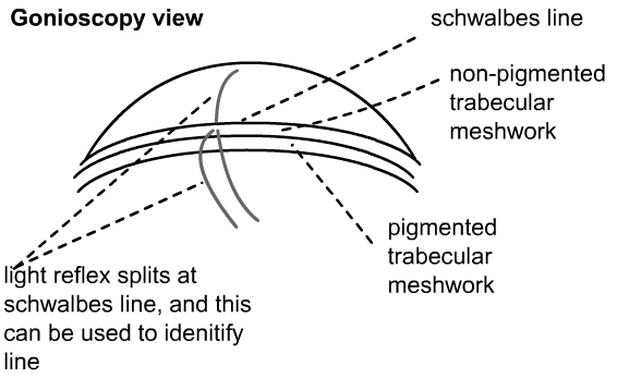
How to find schwalbe's line etc
goniocopy view...double slit shows schwalbe's line


NA-AION & NA-PION
  • non-arteritic anterior ischaemic optic neuropathy
  • ~60y age, always >40y
  • painless loss of vision, like a smudge or smear
  • one or several steps of visual loss
  • some recover vision
  • swollen disc at diagnosis...small optic nerve...'crowded disc'
  • colour vision ~ visual acuity
  • risk factors...diabetes, blood pressure, cholesterol, smoking, high homocysteine level
  • ?nocturnal hypotension
  • if gets worse...check for tumour etc
  • 15%..second eye affected  (if risk factors are treated)
  • posterior ischaemic optic neuropathy (non-arteritic) is related to bleeding, major surgery, hypotension. Usually some risk factors.
  • smaller discs are affected....much less common with large optic disc diameters.


Is visual field loss from glaucoma or toxic optic neuropathy
  glaucoma toxic
field progressive also progressive
cupped? cupping variable
vision normal reduced, dyschormatopsia
colour normal reduced
field arcuate non- central central/paracentral scotoma
    later atrophic
    may be bitemporal
    may improve with treatment


Glaucoma or progressive optic nerve lesion?
  • occupational/drug/food history
  • eg red snapper fish 5x week for years has lots of mercury
  • family history
  • FBC AND B12 and folate and syphilis test
  • lebers...test mitochondria DNA
  • loss of visual acuity ..very rare in (non-advanced) glaucoma
  • field..vertical defects...must exclude CNS lesion
  • rapid...must exclude CNS lesion.
  • if the disc defect is not proportional to field defect... ...must exclude CNS lesion
  • often need MRI to exclude CNS lesion
  • pale rim...must exclude CNS lesion
  • ethanol, methanol, amiodarone, lead, Hg, etc
  • viagra (disputed)
  • endemic Cuba (?cassava), Hungary (?Hg)
  • nutritional B12, thiamine, folate...treat (and treat alcohol/smoking/poor diet amblyopia) with replacement
  • glaucoma does not cause loss in colour vision ...acuity loss always occurs first
  • APD...if present, glaucoma less likely


Glaucoma and night (notes from College, 2007)
  • IOP varies at night
  • lying supine/face down increases the eye pressure ~7mmHg
  • normal blood pressure dips at night...it is not know whether this influences glaucoma
  • Prof Shah feels that patients with Low/Normal tension glaucoma, who also use betablockers, may notice extra dips at night, and these may contribute to reducing optic disc perfusion and increasing visual field loss. Betablockers should be stopped if field is deteriorating (maximum glaucoma therapy), or certainly changed to alternative drugs, if at all possible.
  • dips at night may contribute to AION/RVOs/MIs/RAO etc


Glaucoma and steroids (notes from College, 2007)
  • steroids..tablets, drops, nasal sprays, facial creams cause an increase in IOP
  • this is reversible initially
  • later irreversible
  • eyes with glaucoma/older patients/diabetics/connective tissue disease/ notice bigger increases
  • dose is cumulative
  • dexamethasone is the most potent...2.2x prednisolone 1%
  • IVT..the second injection has a much higher IOP effect
  • IVT causes more increase in younger patients


Glaucoma / cupped discs (notes from College, 2007)
  • a cupped disc can also be due to AION/GCA/optic nerve compression/atherosclerosis/congenital/dominant optic atrophy/previous optic neuritis (Rebolleda 2009)
  • pale rim...glaucoma less likely...usually non-glaucomatous


Glaucoma and arcuate scotomas (notes from College, 2007)
  • these may be caused by optic disc drusen, which may be buried
  • there may be a papilloedema appearance
  • occasionally there is another cause...infiltration of optic nerve/demyelination/aneurysm etc see


Glaucoma and optic disc drusen (notes from College, 2007)
  • may be buried, seen with B scan
  • can differentiate from papilloedema with B scan
  • familial...dominant
  • associated with small optic discs..these disrupt axonal transport...and this causes the drusen
  • associated with short axial length
  • associated with RP/Astreak/PXE/Allaire/Alstrom
  • irregular margins
  • unusual branching of vessels, such as trifurcation, cilioretinal vessel
  • see photos
  • enlarged scotomas
  • superficial drusen..may cause a visual field defect
  • they autofluosce...take photo using he FFA settings without the fluorescein injection
  • papilloedema....hyperaemic/no venous pulsation
  • linked to NAION (NAION may start with transient loss of VA)


Glaucoma notch (notes from College, 2007)
  • notch must be related to the field defect, otherwise suspect another cause
  • see


Asymmetric glaucoma
Suspect carotid artery stenosis


More about pressures and anticardiolipin antibody (includes notes from College, 2008)
  • need <18 at 100% visits
  • target 14mmHg or less
  • each 1mm = 10% progression (Early Manifest Glaucoma Trial)
  • big swings...surgery..suspect poor compliance
  • optic nerve perfusion is related to pressure
  • try and predict progression rate and tell patient
  • Ocular hypertension study...treatment worthwhile if moderate or high risk of POAG
  • Canadian Glaucoma Study: Patients with abnormal anticardiolipin antibody levels and increasing age had faster visual field change. Modest IOP reduction in progressing patients significantly ameliorated the rate of visual field decline.


Blood pressure & ocular perfusion,

Blood pressure may have a role in glaucoma. Some papers indicate that too low a blood pressure may be harmful, whilst others suggest that a reasonably low blood pressure might help.

It is likely that the ocular blood flow (i.e. the blood flow into the optic nerve) is likely to be the most important factor (Costa 09,   Liang 08)

  • excess blood pressure dips at night are likely to be harmful.
  • these dips are likely to be more common in patients with circulation problems, such as migraine or Raynauds (cold hands), and these may contribute to low tension glaucoma
  • a reasonable night time dip is physiological and not harmful
  • too much medication might be harmful, and may be calcium channel blockers may not be the best drug.
  • an ocular perfusion pressure of 50 may be best
  • too high a blood pressure will contribute to retinal vein occlusion
  • see
  • pseuodexfoliation is linked to peripheral vascular disease Eye 2011
  • Similarly, sleeping in the head up postiion may lower IOP 2010


Symptoms (notes from College, 2008)
  • field defects ...unable to read for pleasure
  • reduced hand-eye coordination
  • 33% of glaucoma patients have scotomas


Examining Fields (2008)
  • F  fidelity
  • I   ?? ..type of defect
  • E  extent
  • L  location
  • D  deviation
  • fidelity....another word for reliabiliaty...how many false positives/negatives
  • type of defect..bitemporal...?pituitary
  • extent ...how large the defect is
  • location...where is the defect
  • deviation...is there a pattern deviation that excludes a cataract

Field changes fluctuate BJO 2011


Measure discs (2008)
  • with an HRT!
  • 90d lens...magification 1.4, so a measured 1mm disc is really 1.4
  • 66d lens...measures size  = actual size
  • 90d = 1.33 x disc measurement = actual size
  • 60 = 0.88    x disc measurement = actual size     
  • 78 = 1.2     x disc measurement = actual size
  • average disc = 1.9 mm vertical x 1.8 horizontal
  • peripapillary atrophy is weakly linked to glaucoma .
    • beta..ß.. inner atrophy
    • alpha..α = outer zone atrophy
  • the nerve fibre layer is reduced in thickness in myopia....is this due to glaucoma? BJO 2011. There is a very large range or 'normal' NFL thickness in myopia

types of cupping

  • notch
  • thin rim
  • saucer
  • concentric...even rim
  • see


Notes about fields/disc haemorrhages (2008)
  • 10.2 humphrey often best for advanced glaucoma
  • 24.2 probably best for general use, occasionally need both
  • 30.2 has too many peripheral artifacts
  • ignore fixation losses unless high
  • mention pupil size
  • 5.2 for central field ...eg macular for hydroxycholoraquine toxicity
  • IOHT...measure fields ~1-2 yearly
  • stable glaucoma 1yearly
  • mention disc size when discussing cupping
  • deterioration is related to prognosis see 09
  • loss of field in both hemifields is more likely to lead to progression (eg superior & inferior)  (De Moraes 2009)
  • more aggressive treatment is needed if their is an optic disc haemorrhage (Prata 2009)
  • progression rates average
    • NTG -0.4db/year (normal/low tension)
    • POAG equivalent (high tension) -1.3db/year
    • PEXG -3db/year (pseudoexfoliation)


Glaucoma genes
  • myocilin Gly367Arg mutation....Swiss family, open angle
  • mitochondrial damage
  • review Eye 2012 ..myocillin and optineurin are the prime genes.
  • Eye 2012
    • Risk factors
      • black race
      • untreated blood pressure
      • current smoking
      • family history of glaucoma
      • diabetes
      • myopia
      • central  corneal thickness
    • relatives 22% risk
    • 20 loci
    • MYOC  , JOAG and adult POAG..mitochondrial membrane calcium channel
    • OPTN ..NTG
    • WDR36 & GLCIG


Poor compliance

See Lacey J, Cate H, Broadway DC 2009

  • poor education,
  • lack of faith in efficacy
  • difficult using drops
  • forgetting
  • practical problems
  • older age...multiple problems
  • younger..non-acceptance


Chronic conjunctivitis

We are all familiar with a true allergy (eczema around the eyes, itchy eyes), but many patients develop more chronic changes.

  • the conjunctiva may be red
  • there may be epiphora (watery eye)
  • there may be a papillary conjunctivitis, especially in the lower fornix.
  • it is difficult to determine whether the problem is due to the drug in the eye drops or the preservative; preservative free glaucoma eye drops may help.

allergic conjunctivitis

allergic conjunctivitis: lots of papillae


SLT laser

SLT lowers pressure Singh09

  • it lowers pressure about 6mmHg
  • using prostaglandin inhibitors at the same time does not lower the pressure further
  • the effect starts to wear off at 6 months, with little effect at 1 year.
  • www.brendanmoriarty.com/glaucoma/slt/  
  • Moriarty Laser Treatment leaflet
  • few laser burns are targeted at the trabecular meshwork. This laser is carried out in outpatients and very safe, with few risks. Patients can go home a few minutes after the laser, though occasionally the pressure needs to be tested one hour later. Both eyes are treated at the same session.
  • The angle must be open, that is 'open angle chronic glaucoma', not closed.
  • In Birmingham this is only available at the Birmingham and Midland Eye Centre.
  • Technically the outflow drainage is increased after 3600 laser from 0.11μl/minute/mmHg to 0.5, lowering pressure  from 26 to 12mmHg.
  • BJO 2010


slt laser is a new  treatment for open angle glaucoma


The address of this site ('org' changing to 'nhs') is changing from http://www.goodhope.org.uk/departments/eyedept/ to http://www.goodhope.nhs.uk/departments/eyedept/
Eye website feedback -- Heartlands -- page edited October 2011 -- Public transport to Good Hope --