| Vitreo-macular
traction (VMT) and Case 24 David Kinshuck |
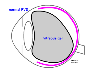
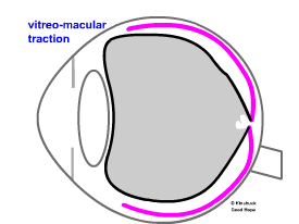
In this condition the vitreous gel shrinks and detaches from the back of the eye (the retina), a bit like a regular PVD as opposite (posterior vitreous detachemnt). A regular PVD is more or less a normal aging change. But unlike a PVD, the vitreous gel remains stuck to the macula in the centre, and pulls it. This may cause some distortion of central vision. But the 'VMT' is transient. There is a 90% chance of a complete pvd (posterior vitreous detachment) developing, with stable good vision. See VMT animation. However, there is also a ~10% chance of developing a complete macular hole, with some loss of sight (surgery may be needed). This occurs when the vitreous pulls the retina in the foveal area to the extent that it creates a hole. The hole may cause loss of vision, and may be helped with surgery. The cause of the VMT is linked to PVD formation, when the vitreous gel shrinks. Normally the gel separates completely from the retina, but in this condition the back face of the gel remains stuck to the retina, in the foveal area. If the distortion persists and is really annoying then vitrectomy can help.
|
a normal 'PVD' ..the vitreous detaches without pulling any of the retina, The space behind the vitreous becomes with a fluid rather than a gel.
VMT..the vitreous has detachched and is pulling the macula area of the retina in the centre. |
Case 24 VMT ..vitreo-macular traction
Not treatment was needed and the patients maintainded good vision. See the scan below. |
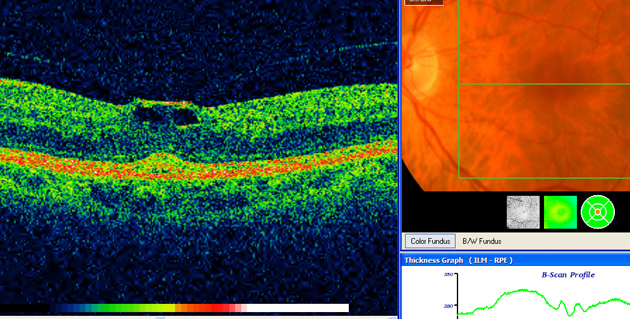
| Vitreo-macular traction with a small
foveal cyst |
|
| The address of this site ('org' changing to 'nhs') is changing from http://www.goodhope.org.uk/departments/eyedept/ to http://www.goodhope.nhs.uk/departments/eyedept/ |
|