====
Good Hope Hospital Eye Clinic

Low Tension/Normal Pressure Glaucoma
David Kinshuck


What is glaucoma?

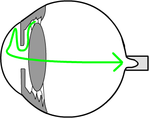
Glaucoma is a condition in which the nerve at the back of the eye is damaged. If severe, it can cause blindness, but most people do keep good sight indefinitely.

It is explained in more detail on the Glaucoma page, which should be read in addition to this page.

In ordinary glaucoma the nerve is damaged by an unusually high pressure in the eye. The high pressure stops the blood entering the tiny blood vessels in the part of the optic nerve that enters the eye.

Animation


a blocked drain causes the eye pressure to rise and damage the optic nerve

In ordinary glaucoma the nerve is damaged by pressure from a build up of pressure inside the eye
enlarge


Low tension glaucoma

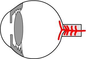
In this type of glaucoma we believe not enough blood reaches the tiny arteries in the optic nerve.
Blood from the heart flows towards the head, and then to the eye. Behind the eye the blood flows in a small artery, and just behind the eye the small blood artery enters the optic nerve itself.
Inside the nerve this small artery provides even smaller tiny branches that reach the tissues of the nerve.
Without its blood supply, the nerve becomes damaged, and the sight will be reduced. An alternative explanation is that there is a fault with the blood vessel oxygen extraction process.

See an optic nerve circulation animation

In low tension glaucoma it is thought the primary problem is too low a pressure in the blood supply to the nerve. This is rather like having a low water pressure in a house... not enough water comes out of the tap. 
We do no know why this is, but only rarely does the nerve become badly damaged.

Treatment is drops in most patients. The drops lower the pressure in the eye so more blood can flow in. Occasionally an operation is needed as below.

low tension glaucoma may be cuased by poor blood flow to the optic nerve

In low tension glaucoma not enough blood reaches the optic nerve (the tiny blood vessels supplying the optic nerve are shown in red).

enlarge


How does the treatment work?

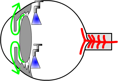
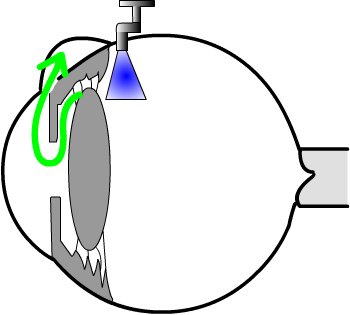
Normally, fluid (a little like water, called ‘aqueous humour’) is made in the middle chamber of the eye.

It then flows through the pupil to the front chamber, and then out of the eye through small drainage channels.

These channels are like a 'sieve' in the kitchen used to drain rice.


aqueous fluid produced in the eye drains out; blood flows into the optic nerve

Aqueous fluid (green) is made in the middle chamber of the eye (shown by the 'taps'), and drains out at the side of the front chamber.
Blood from the heart originally enters the nerve (shown in red).
enlarge


Eye drops

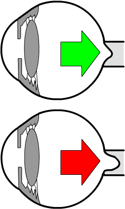
Eye drops containing drugs can ‘switch the tap’ off that makes the fluid (top half of eye opposite). Less fluid in the eye leads to a lower less pressure (like a tyre that is not pumped up as much). This allows more blood to enter the eye, and more blood flowing to supply the optic nerve.

Other drops open up the drain that lets the fluid out (bottom half of eye opposite)

drops either switch off the tap or increase the drainage

Less fluid is made (top half), or more fluid drains out of the eye. Both make the eye is softer and this lets more blood flow in to supply the nerve. enlarge


An Operation

Occasionally an operation may be necessary. As part of the operation a small hole is made to let more fluid out of the eye. Your ophthalmic surgeon will discuss the pros and cons of the operation if it is necessary.

trabeculectomy...surgery is occasionally needed to lower eye pressure

A small hole is made at the top of the eye, under the eyelid, to let the fluid drain out of the eye. As a result, the eye becomes softer, and more blood can flow in.
enlarge


How is your sight affected?


Usually the sight is not greatly affected in this condition. In younger patients (30-60y), the sight may get worse over many years. This can cause variable sight that changes during the day. Alternatively, the sight may be patchy, with clear areas and fuzzy areas.

The damage can be similar to that of ordinary chronic glaucoma, as opposite.

At first the sight is normal,


then a small area of poor vision may develop.


This can extend, affecting much of of the sight,


or nearly all the sight.

visual fields get gradually worse in untresated chronic glaucoma


Visual fields

The sight is tested in the clinic for this type of patchy vision, by a visual field testSee a result.

In this test, you have to put your head in a type of 'gold-fish bowl'. Lights are flashed on, and you have to press a button when you see the lights. The computer in the machine compares your result to that of a 'normal' person of your age, and prints out a serious of dots and black areas. The black areas are the area of damaged sight.


Can you become blind?
Only if you were very unlucky will your sight become very poor. Hardly anyone becomes blind in that they cannot see anything at all, and only occasionally will your sight become very bad.

If an ophthalmic surgeon recommends the drops, you should use them (unless you get side effects). Often the drops can make all the difference between keeping your sight and loosing it. (The same applies to an operation if your ophthalmic surgeon advises you strongly you should have one).

Surprisingly, only two out of three patients recommended drops actually use them regularly. If a doctor has advised to use the drops and you choose not to, it is only fair to be honest and mention this to your doctor.
For most people with this type of glaucoma it makes very little difference if you miss the drops now and again, so do not worry if you forget to use them now and again.


What tests do you need in the eye clinic?
  • visits to the clinic every 12 months if the pressure is controlled

  • 3 monthly or so if the pressure is higher and you are trying to get it lower

  • a pressure test each visit: ideally the pressure is about 12mmHg for younger patients

  • a visual field test every 6-12 months, to test for patchy vision  See.

  • an examination of the optic nerve each visit: the doctor looks onto your eye and can see the nerve. The nerve looks ‘deeper’ if it gets damaged.


A healthy optic nerve

a cupped optic nerve...caused by high pressure/too much fluid in the eye

A cupped, or damaged,
optic nerve

enlarge


What can you do to help?

See General health below. The main thing is to use drops if your doctor has advised you to.

A few people have low blood pressure at night; ask you general practitioner if this is likely. There are no tablets and there is no magic cure though.

Strangely, people with this type of glaucoma are more likely to have migraine or poor circulation in their hands (doctors call this 'Raynauds').

A low blood pressure is helpful (Kim 2010). Low tension glaucoma is related to general health vascular problems (Leung 2010).


Your general health in glaucoma/intraocular hypertension

Remember that your general health and lifestyle have a major impact on glaucoma and your sight. BJO 12

  • Smoking does increase the eye pressure, and will make your glaucoma worse. It also increases the risk of retinal vein occlusion (which occur in glaucoma), macular degeneration, cataracts, stroke, and heart attacks. The exact risk is not known, but in some patients 20 ciagettes / day will increase loss of vision by 400%. Passive smoking 20 a day is equivalent to smoking 5 cigarettes a  day, and increase visual loss 100%.

  • metabolic sydrome is strongly related intraocular pressure (Nature 2010). This is related to (as below) lack of exercise, high blood pressure, obesity and liver and kidney problems. Treatment of the metabolic syndrome will help lower eye pressure 0.8mmHg..

  • Exercise lowers the eye pressure, 'an hour a day (walking) will keep the doctor away', and half an hour will help a lot. See. Any exercise will do... eg walking, swimming, gardening.

  • high blood pressure will be harmful as this may, together with the glaucoma, cause a retinal vein occlusion. A blood pressure of 140 systolic or less, may be best, and may be lower the better as long as you feel well. See   But there are other views. We now know that eye pressure and blood pressure are related; the implication of this is that lowering blood pressure will help in the treatment of glaucoma. So keeping your blood pressure reaonably low is an important part of the treatment, although too much medication may cause problems.

  • Obesity results in high blood pressure, lack of exercise, and indirectly will cause problems. Many obese patients suffer from sleep apnoea....

  • Oily fish will help the circulation and probably help prevent retinal vien occlusions. Red meat  (including pork/ham) increase blood pressure; they are best replaced, at least in part, by fish, and pulses such as lentils and beans. Nuts have some healthy fats, and may be helpful in small amounts...but they are 'fattening' and help to put weight on. 

  • 9 portions of vegetables or fruit a day will also help to keep your retinal veins and macula healthy, and prevent cataract formation...9 a day (men) 7 (women) keeps the doctor away...

  • A high saturated fat diet is very harmful as it will contribute to many conditions. A balanced diet is recommended. Saturated fat comes from full-fat diary products such as milk, cheese, many cakes and biscuits, and red meat.

  • Salt will increase blood pressure.

  • binge drinking, e.g. 4 pints of beer in one day, or more that 3 glasses of wine/day, will cause a rise in blood pressure, again contributing to all these conditions.

  • Sleep problems. New research links sleep apneoa with glaucoma. Sleep problems are very common in obesity...these are likely if you snore a lot. Logically if you are overweight, it is important to lose weight to help your glaucoma. So if you do snore a lot, do lose weight, and ask your doctor if you need to be tested for sleep apnoea.

  • Steroid medication puts eye pressure up. Even inhaled nasal steroids can increase the pressure 2.5mmHg. If you use nasal steroids (eg for allregic rhinitis), ask your doctor if you can reduce or stop them. Steroid tablets certainly may put eye pressure up; ask your doctor if you can reduce the dose (Do not reduce steroid tablets without seeking advice.).

  • Retinal vein occlusions: patients with glaucoma are much more prone to retinal vein occlusion. The risk of a vein occlusion is reduced by control of the glaucoma, blood pressure, and all the other general health risk factors immediately above.


CNS changes

There may be CNS changes in LTG. RCO 2011.


Leaflet download

this leaflet

This web page/leaflet is designed to accompany the Glaucoma page or Glaucoma leaflets available in an Eye Clinic; ask for one in your clinic if you have not seen one.

a leaflet explaining low tension glaucoma


Notes for professionals
Need careful history
  1. test colour vision
  2. check APD
  3. check disc pallour relates to field
  4. look for notch & nerve fibre layer defect and relationship to field
  5. patients should not notice defect..if patients notices defect, risk of neurological problem higher (I would add particularly observant patients may notice glaucoma defects.)
  6. hyper-homocyseinaemia (MTHFR gene) is linked to NTG
  7. CSF pressure may be relevant.
  8. vitamin B12 an folate tested needed in all patients, perhaps every 5 years


The address of this site ('org' changing to 'nhs') is changing from http://www.goodhope.org.uk/departments/eyedept/ to http://www.goodhope.nhs.uk/departments/eyedept/
Eye website feedback -- Heartlands -- page edited October 2011 -- Public transport to Good Hope --