Understanding Macular Disease David Kinshuck |
|
|
|
Introduction |
|
There are many types of macular disease. The commonest type is age-related macular degeneration (ARMD) is discussed here. Macular disease also occurs in Diabetic maculopathy is not discussed here in detail but many of the principles do apply. All types of serious macular disease affect the central vision, making it difficult to read, watch TV, or see faces. This page explains macular disease in general, with some details about age-related macular degeneration in particular. See the page Poor vision-magnification for magnifying aids, and the page Hints & Coping for other advice about how to cope. The text may appear incomplete unless you read all the three pages. These pages were written by David Kinshuck, Jayne Kempster, & Bruce Fisher, with help from Trish Cooper. Contact the RNIB or the United States NIH for more advice. This site is excellent www.macula.org |
|
What is the retina? .....the eye as a camera |
|
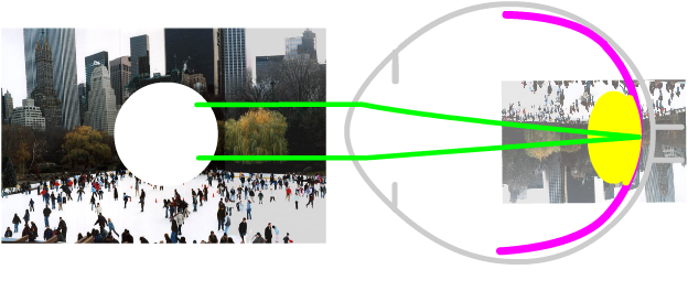
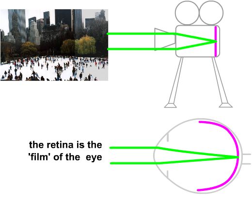
Thinking of a camera can help you understand macular
disease. A camera lens focuses a picture onto a film
inside the camera. In our eyes a similar thing happens,
but the film is replaced by the retina.
The retina 'makes' the pictures of the world that we see, converting the light into electrical signals that are then sent on to the brain. |
 |
|
The Macula |
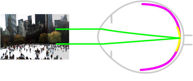
| The central area of the retina is called the macula. The macula is
special, as it is the the most sensitive part of the retina. It makes
out the fine details of the things we look at, peoples' faces, bus
numbers, reading and writing, and the letters on an optometrists chart.
Whenever we look at an object, the image focuses on the macula. If the macula is damaged all these things we see in fine detail are misty. The picture is still there but we cannot make out any of the detail. The brain builds up details of what we see by moving the macula over an object.
|
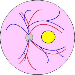
If
a doctor or optometrist looks into the retina can be seen. |
|
Macular disease |
|
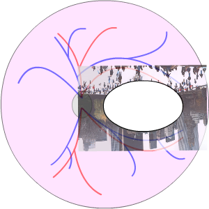
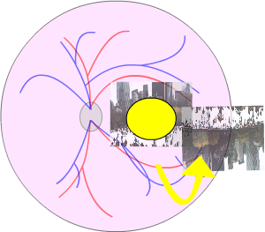
A healthy retina will produce a clear image, like a normal film in a camera. But in macular damage the image will not be clear. For example if the film was scratched in the middle, the 'scratch' would show up in the middle of the photograph like a black mark or smudge of ink, as illustrated below. This is similar to damage caused by macular disease such as age related macular degeneration or diabetic maculopathy... the surrounding retina is not as sensitive as the macula, and cannot detect details such as writing or peoples' faces. Sadly, the damage cannot be repaired. Because the detail in pictures is lost by damage to the macula, no amount of change to your glasses can restore this detail. So people with age related macular degeneration or diabetic maculopathy find it difficult to read, write and or recognise faces. Healthy Macula In a camera a film will detect a detailed image. When
the film is developed this is turned into a clear picture. |
 |
| Macular disease
But if the film of the camera is damaged in some way, no amount of focusing would produce a clear picture. For example, if the film was 'scratched' in the middle, the scratch would show up in the middle of the photograph like a black mark or smudge. This is similar to damage caused by macular disease such as age related macular degeneration, as shown below. |
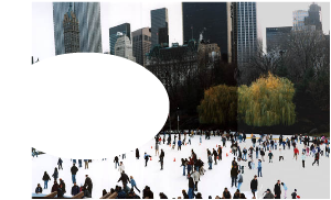
A damaged macula: an image with a central blurred area |
The sight inmacular disease |
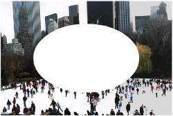
| As above, the macula area of
the retina is responsible for the sharpness and details of what we
see, when it is damaged pictures loose all their detail. Because of
this no amount of change to glasses can restore the sight so details
can be seen normally.
So people with macular degeneration find it difficult to read, write and recognise faces. People with macular degeneration may be able to walk through a busy supermarket, but not be able to see the price tags on the food. Just like the camera if the centre of the film is damaged, it may not be possible to see a person’s face in the centre of a photograph, but an outline of the person and the other people nearby can still be seen. People with macular degeneration may recognise where they are by seeing things and places at the sides. (People without macular degeneration may find this difficult to understand!) .
|
Macular damage, usually due to age related macular degeneration
The central vision is lost in severe macular degeneration
|
|
|
The causes of macular disease |
The commonest cause is ARMD, age-related macular disease. There are other types of macular disease, such as the group of Juvenile Macula Dystrophies, macula holes, and epriretinal membranes. |
|
Will spectacles help in macular disease? |
| New spectacles
may help a little, but it is important to make the
best use of the remaining eyesight.
There are two ways to do this: These two techniques are used when people with ARMD try to read. Also see hints and Low Vison Clinic. |
|
Making maximum use of side vision |
| To make the most of the peripheral
vision ….the side vision…. you need to hold your head in
an unusual position to so that whatever you are looking at is imaged
(in the eye itself) at the side of the macula, under, or above it,
on the healthy area of the retina. Each person needs to experiment to find the best viewing angle. Most people who have macular degeneration find that they automatically do this. It is unusual for this technique to be taught, it is solely a matter of moving the head to obtain the best possible vision. This technique occurs naturally as the disease progresses. It may be necessary to learn the technique when looking at details, such as peoples’ faces. |
Looking sideways can bring people or objects or words into view.
By looking to one side the image
on the retina moves from the damaged macula on to the healthy
retina. |
|
Magnification |
|
Magnifying an object or words being read seems the obvious answer: if the object is bigger it can be seen by the part of the retina outside the damaged area. Remember the person who can see to get about in the supermarket… the sight at the sides is normal. There is a problem. When looking through a magnifier the field of view is small, so only a few words can be seen at one time. To overcome this you need to move the magnifier along the line to ‘piece the sentence’ together. This takes a lot of practice: you have to move your eyes along the line, whilst trying to use the side vision, whilst looking through a magnifying glass, trying to guess missing sections of words. Reading is still possible even with medium size print, although it is usually very slow. |
Reading is difficult with severe macular degeneration, but is slightly easier magnifying the writing and looking to one side. |
|
|
Hallucinations are common if you have very poor sight: the Charles Bonnet syndrome |
| Many people with
poor sight notice hallucinations. Such hallucinations are very common
in macular degeneration. These can take various forms, and are described
in more details in the BMJ
here, and here. If you see such things that are not really
there, you are not going mad...it is quite normal if you have poor sight.
If they are very disturbing discuss them with your doctor or ophthalmologist. |
www.geocities.com/ allspiderwomen/galspidr.htm |
For researchers: visual function questionaire |
| http://www.nei.nih.gov/resources/visionfunction/vfq_ia.pdf |
| The address of this site ('org' changing to 'nhs') is changing from http://www.goodhope.org.uk/departments/eyedept/ to http://www.goodhope.nhs.uk/departments/eyedept/ |
|